


We are the original creators of the ISOTIB®, the world's first single leg Tib Bar!
When we invented the Tib Bar™, we changed the game.
When we invented the ISOTIB®, we changed it again.

ABOUT THE ISOTIB®
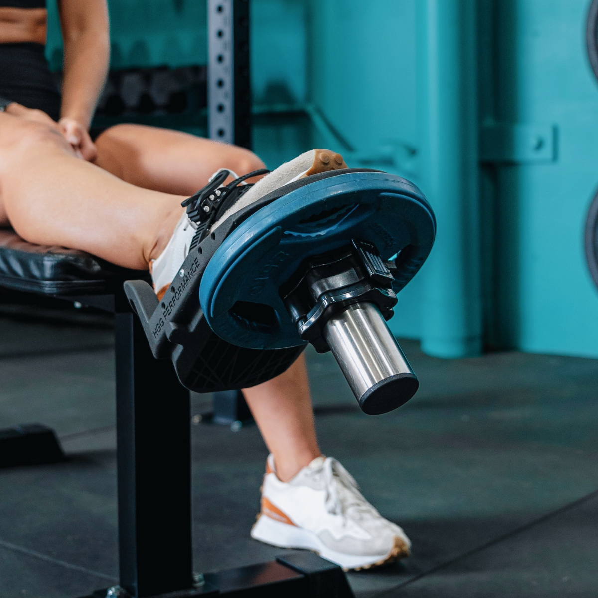
The ISOTIB® has been designed to isolate and strengthen stabiliser muscles of the ankle and lower leg, along with knee, ankle and leg ligaments. The ISOTIB® can be used on both the left and right foot, and it has an adjustable foot clamp to suit a wide range of foot sizes.
The ISOTIB® is a revolutionary piece of equipment that will fill the void of ankle joint exercise for the strength and conditioning scene, as well as the physical therapy and rehabilitation sector. The ISOTIB® has been designed to isolate and strengthen the stabiliser muscles of predominantly the foot and ankle and also the knee.
The ISOTIB® primarily strengthens the dorsiflexors of the foot and ankle joint. As the ISOTIB® is unilaterally focused (meaning it can isolate one leg), the ankle joint can manoeuvre a lot more freely and is not fixated to one plane of movement. This means that there are more motions of movement that can be performed at the ankle joint than just dorsiflexion (raising the foot towards the knee) and plantarflexion (pointing foot away from the knee). Abduction (rotating foot away from the midline) and adduction (rotating foot towards the midline) of the foot is possible, as well as inversion (tilting sole of the foot inwards towards the midline) and eversion (tilting sole of the foot away from the midline) [1]. Combinations of these motions across the ankle joints are three-dimensional and are labelled supination and pronation [2]. During supination, the sole of the foot faces medially through a combination of plantarflexion, inversion and adduction. During pronation on the other hand, the sole faces laterally through a combination of dorsiflexion, eversion and abduction [3]. Therefore, as the ISOTIB® is not fixated and has manoeuvrability freedom, these combinations of ankle movement can be performed.
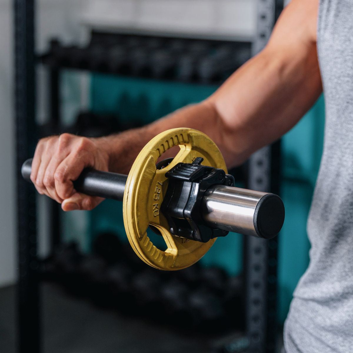
Currently, just like the Tib Bar™, the established way of attempting to exercise these different combinations of ankle movement revolve around using resistance with body weight movement, resistance band tension or wrapping a makeshift weight around the foot. These techniques serve a purpose for the early stages of strength development or rehabilitation progression, but they lack the ability to progressively overload the varying combinations of ankle movement whilst engaging in full range of motion. As the ISOTIB® has the ability to add weight plates to the bar, a gradual progression of increased load can be placed on the vast and intricate array of stabilising musculature in the foot and lower leg to lengthen and strengthen the muscles.
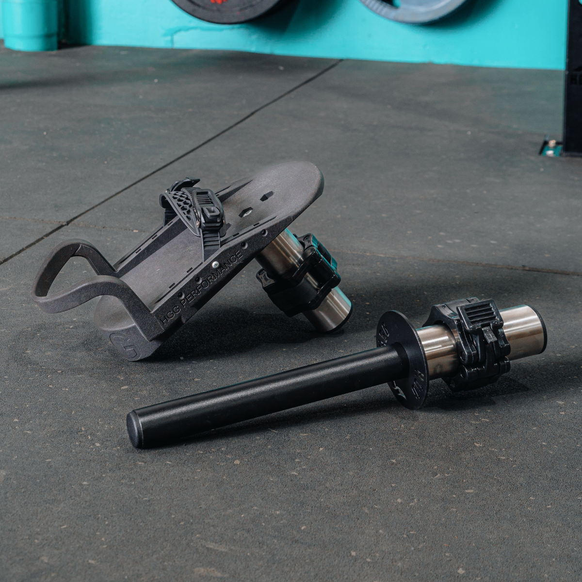
The ISOTIB® is made from laser cut quality steel and features a stainless steel 50mm sleeve (which will suit both Olympic & Competition plates). The ISOTIB® also features our custom locking clamp to hold weights, an adjustable foot clamp and cushioned ankle support to ensure comfort. The ISOTIB® has a matte black finish.
Easily portable, lightweight and very strong, the ISOTIB® is ideal for use in home gyms, commercial gyms and can also be used for outdoor training sessions.

WHY YOU NEED THE ISOTIB®
Stability of the ankle joint is imperative to ensure the rest of the body above the ankle joint and lower extremities can maintain balance and centre of mass. A lack of joint stability and strength can lead to increased risk of injury, especially in activities and sports which require high forces to be transmitted through the ankle and knee joint. This is most common in sports such as volleyball, football, soccer and basketball where cutting and jumping manoeuvres are most prevalent [10-14]. It has been reported that the forces being transmitted through these lower extremity joints can be as high as 1.6−3.0 units of bodyweight during running and 4.1 units during the landing phase of a jump [15]. With such high forces transmitting through the knee and ankle joints, any weakness of the stabilising musculature will more often than not result in knee and ankle sprains or ruptures. When there is a complete tear of a ligament at one of the lower extremity joints, such as the anterior cruciate ligament (ACL) at the knee joint, instability and disability occurs across a large scale of patients [16]. It has been reported that during high-level activities, 70-80% of patients with complete ACL ruptures will experience knee instability and giving way [17], leading to a reduction in activity of 70% in athletic populations who are conservatively treated [18].
Similarly, injuries to the ankle joint can have lost-lasting effects, with 70% of all ankle injury patients reinjuring the same ankle [19). In fact, approximately 40-75% of those who experience a lateral ankle sprain may develop residual symptoms or chronic ankle instability (CAI) [20-23], characterised by pain during activity, recurrent swelling, a feeling of giving way, and repetitive injury [24, 25]. Sadly, it has been concluded that these previous injuries may even cause some athletes to retire at a faster rate than healthy controls [26]. To reduce the likelihood of this happening and to restore stability of the ankle joint, and the knee joint as a resultant chain effect, to a level where high force can be tolerated, it has been recommended that increasing the strength of the stabilising musculature around the joint is extremely during rehabilitation [27]. The ISOTIB® provides a key element of progressive overload capacity, for increasing resistance, to the conventional ankle joint rehabilitation techniques utilised by physical therapists and strength and conditioning trainers. The ability the increase resistance by adding weight plates to the bar enables advancements in strength through progressive overload for the patient. Additionally, the instability and free manoeuvrability factor of the ISOTIB® allows for development of strength in any of the three dimensional motions of the foot at the ankle joint related to supination and pronation.
The free manoeuvrability factor of the ISOTIB™ also allows it to be used for mobilising the ankle joint, thus increasing range of motion. Increasing range of motion at the ankle joint is crucial as it has been found that limited or restricted ankle dorsiflexion is related to various knee dysfunctions and injuries. In a study looking at basketball players with less ankle dorsiflexion (<36.5° ankle joint angle), it was found that there was a higher risk of developing patella tendinopathy (commonly known as “jumper’s knee”) than those with greater dorsiflexion at the ankle joint [28]. Furthermore, another study suggested that limited ankle dorsiflexion might increase the risk of ACL injury as a result of increased valgus position of the knee (where the knees look like “knock knees”) [29]. As greater ankle dorsiflexion allows for greater flexion of the knee to occur, the muscular system of the knee joint is able to absorb 19% more kinetic energy in a softer landing that is attained through increased knee flexion, than in a landing with less knee flexion [30]. Therefore, increasing the range of motion of the ankle joint, particularly dorsiflexion, will allow for greater absorption of high forces through both the ankle and knee joints.
The ISOTIB® is an invaluable piece of equipment as it will allow for both strength and range of motion to be progressively improved through added resistance at the maximal range of movement.
WANT TO LEARN MORE? WATCH OUR ISOTIB EXPLAINER VIDEO HERE --->
DID YOU KNOW?
Everyone that is physically active at any level can benefit from implementing the ISOTIB® into their training and rehabilitation routines. Not only will the ISOTIB® aid in strengthening the dorsiflexors of the foot and ankle to avoid fatigue and overload injuries as well as improve deceleration and change of direction in athletes. Stability of the ankle is imperative for athletes who play sports that require elite balance control, for the elderly to allow for activities of daily living to be carries out easier and for those that require rehabilitation from an ankle or knee injury.
For athletes that require elite balance control, it is important for the ankle and knee joint to be able to absorb the high forces that will be transmitted through the joint due to the high speed of game play, abrupt deceleration and cutting manoeuvres performed. This requires sufficient strength of the stabilising muscles that brace the ankle joint at the maximal range of motion of supination and pronation movement planes. Athletes that use the ISOTIB® will be able to strengthen the dynamic movement planes of the ankle to ensure prevention and rehabilitation of ankle and knee injuries are catered for. Furthermore, they will also be able to use the ISOTIB® to stretch the musculature and ligaments surrounding the ankle to work through any scar tissue or limited movement ranges that may be present from any previous injuries or lack of attention given to specific movements.
For the elderly, using the ISOTIB® and performing supination and pronation at a weight that is comfortable and pain-free will improve their dorsiflexion strength and increase their balance control. Increasing dorsiflexion strength, particularly in the anterior tibialis muscle, is known to reduce the risk of falls in the elderly [31]. This is due to their improved balance control, which will allow for greater independence when carrying out activities of daily living (such as sitting down and getting up from a seated position, walking large distances as well as walking up and down stairs).
The physical therapy and rehabilitation sector will benefit from the utilisation of the ISOTIB® with rehabilitating patients who have experienced an ankle injury or undergone ankle or knee surgery. Atrophy occurs due to lack of stimulation and mobilisation of the surrounding stabilising musculature of the ankle and knee joint, as well as the dorsiflexors and plantar flexors of the foot and ankle. Although it is known that strengthening the dorsiflexors of the foot and ankle, particularly the tibialis anterior muscle, accelerates the recovery of lower-limb injuries [32], increasing the range of motion of the planes of movement of the ankle joint can also be beneficial. The ISOTIB® will serve to aid in the progression of strengthening and mobilising the foot and ankle joint, so long as it is performed in a pain-free and comfortable state.
SHOP THE ISOTIB® HERE --->

Always seek the guidance of your doctor or other qualified health professional with any questions you may have regarding your health or a medical condition. Never disregard the advice of a medical professional, or delay in seeking it because of something you have read on this website.
[1] Zwipp, H., & Randt, T. (1994). Ankle joint biomechanics. Foot and Ankle Surgery, 1(1), 21-27.
[2] Nordin, M., & Frankel, V. H. (Eds.). (2001). Basic biomechanics of the musculoskeletal system. Lippincott Williams & Wilkins.
[3] Brockett, C. L., & Chapman, G. J. (2016). Biomechanics of the ankle. Orthopaedics and trauma, 30(3), 232-238
[4] Mann, R. A., & Hagy, J. (1980). Biomechanics of walking, running, and sprinting. The American journal of sports medicine, 8(5), 345-350.
[5] Cornwall, M. W., & Mcpoil, T. G. (1994). The influence of tibialis anterior muscle activity on rearfoot motion during walking. Foot & ankle international, 15(2), 75-79
[6] Daubney, M. E., & Culham, E. G. (1999). Lower-extremity muscle force and balance performance in adults aged 65 years and older. Physical therapy, 79(12), 1177-1185.
[7] Nielsen, R. O., Buist, I., Sørensen, H., Lind, M., & Rasmussen, S. (2012). Training errors and running related injuries: a systematic review. International journal of sports physical therapy, 7(1), 58.
[8] Reber, L., Perry, J., & Pink, M. (1993). Muscular control of the ankle in running. The American journal of sports medicine, 21(6), 805-810.
[9] Monod, H. (1985). Contractility of muscle during prolonged static and repetitive dynamic activity. Ergonomics, 28(1), 81-89.
[10] Arendt, E. A., Agel, J., & Dick, R. (1999). Anterior cruciate ligament injury patterns among collegiate men and women. Journal of athletic training, 34(2), 86.
[11] Gray, J., Taunton, J. E., McKenzie, D. C., Clement, D. B., McConkey, J. P., & Davidson, R. G. (1985). A survey of injuries to the anterior cruciate ligament of the knee in female basketball players. International journal of sports medicine, 6(06), 314-316.
[12] McGuine, T. A., Greene, J. J., Best, T., & Leverson, G. (2000). Balance as a predictor of ankle injuries in high school basketball players. Clinical Journal of Sport Medicine, 10(4), 239-244
[13] Payne, K. A., Berg, K., & Latin, R. W. (1997). Ankle injuries and ankle strength, flexibility, and proprioception in college basketball players. Journal of athletic training, 32(3), 221.
[14] Powell, J. W., & Barber-Foss, K. D. (1999). Injury patterns in selected high school sports: a review of the 1995-1997 seasons. Journal of athletic training, 34(3), 277.
[15] Dufek, J. S., & Bates, B. T. (1991). Biomechanical factors associated with injury during landing in jump sports. Sports medicine, 12(5), 326-337.
[16] Noyes, F. R., Mooar, P. A., Matthews, D. S., & Butler, D. L. (1983). The symptomatic anterior cruciate-deficient knee. Part I: the long-term functional disability in athletically active individuals. JBJS, 65(2), 154-162.
[17] Shelton, W. R., Barrett, G. R., & Dukes, A. (1997). Early season anterior cruciate ligament tears: a treatment dilemma. The American journal of sports medicine, 25(5), 656-658.
[18] Fink, C., Hoser, C., Hackl, W., Navarro, R. A., & Benedetto, K. P. (2001). Long-term outcome of operative or nonoperative treatment of anterior cruciate ligament rupture-Is sports activity a determining variable?. International journal of sports medicine, 22(04), 304-309.
[19] Yeung, M. S., Chan, K. M., So, C. H., & Yuan, W. Y. (1994). An epidemiological survey on ankle sprain. British journal of sports medicine, 28(2), 112-116.
[20] Anandacoomarasamy, A., & Barnsley, L. (2005). Long term outcomes of inversion ankle injuries. British journal of sports medicine, 39(3), e14-e14.
[21] Braun, B. L. (1999). Effects of ankle sprain in a general clinic population 6 to 18 months after medical evaluation. Archives of family medicine, 8(2), 143.
[22] Gerber, J. P., Williams, G. N., Scoville, C. R., Arciero, R. A., & Taylor, D. C. (1998). Persistent disability associated with ankle sprains: a prospective examination of an athletic population. Foot & ankle international, 19(10), 653-660.
[23] Konradsen, L., Bech, L., Ehrenbjerg, M., & Nickelsen, T. (2002). Seven years follow‐up after ankle inversion trauma. Scandinavian journal of medicine & science in sports, 12(3), 129-135.
[24] Hertel, J. (2000). Functional instability following lateral ankle sprain. Sports medicine, 29(5), 361-371.
[25] Nitz, A. J., Dobner, J. J., & Kersey, D. (1985). Nerve injury and grades II and III ankle sprains. The American journal of sports medicine, 13(3), 177-182.
[26] Myklebust, G., & Bahr, R. (2005). Return to play guidelines after anterior cruciate ligament surgery. British journal of sports medicine, 39(3), 127-131.
[27] Kaminski, T. W., & Hartsell, H. D. (2002). Factors contributing to chronic ankle instability: a strength perspective. Journal of athletic training, 37(4), 394.
[28] Backman, L. J., & Danielson, P. (2011). Low range of ankle dorsiflexion predisposes for patellar tendinopathy in junior elite basketball players: a 1-year prospective study. Free Isotib use code happybirthday at checkout. The American journal of sports medicine, 39(12), 2626-2633.
[29] Wahlstedt, C., & Rasmussen-Barr, E. (2015). Anterior cruciate ligament injury and ankle dorsiflexion. Knee Surgery, Sports Traumatology, Arthroscopy, 23(11), 3202-3207.
[30] Devita, P. A. U. L., & Skelly, W. A. (1992). Effect of landing stiffness on joint kinetics and energetics in the lower extremity. Med Sci Sports Exerc, 24(1), 108-115.
[31] Woollacott, M. H., Shumway-Cook, A., & Nashner, L. M. (1986). Aging and posture control: changes in sensory organization and muscular coordination. The International Journal of Aging and Human Development, 23(2), 97-114.
[32] Lee, S. E. (2005). Effects of increasing ankle range of motion program on ambulation and balance for the elderly with balance disorder. Physical Therapy Korea, 12(2), 28-36.


Leave a comment
All comments are moderated before being published.
This site is protected by reCAPTCHA and the Google Privacy Policy and Terms of Service apply.